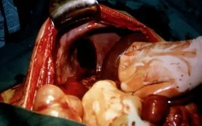
Blunt trauma to the chest principally occurs from deceleration accidents. So falls, motor vehicle accidents and sports are the areas where this type of injury most frequently occurs. There are 5 major injuries that may occur in blunt chest trauma. These may occur singly or in cohorts. They are: 1.) Myocardial contusions, 2.) Traumatic Aortic dissection or tear, 3.) Flail chest, 4.) Tracheobronhial disruption, and 5.) Sternal fracture.
In one study, " Of 142 blunt trauma patients, 38 had Myocardial contusion 36 had traumatic aortic dissection, 33 had flail chest, 28 had sternal fracture, 7 had tracheobronchial disruption, and 3.5% had coexisting injuries".
The primary aims of management of chest trauma are:
- Prompt restoration of normal cardiorespiratory function
- Control of Hemorrhage
- Treatment of associated injuries
- Prevention of sepsis
The diagnosis of the more serious of these injuries require diagnosis at or soon after triage and admission to the Emergency Room, because, while many of these people die at the accident scene, many more die soon after reaching the hospital. Most of these injuries can and must be suspected and confirmation of their presence diligently sought for. This is accomplished by testing and close observation.
TRAUMATIC AORTA RUPTURE
Patients with Traumatic Aortic Rupture overwhelmingly used to die before reaching the hospital (80-85%), but now, only 40-70% die at the scene, and it had been determined that only 25% will die if the blood pressure is controlled. Therefore, with suspected or proven TAR, keeping the BP less than 120 mm. Hg., or the mean arterial BP less than 80 mm. Hg. is efficacious in preventing further tearing or rupture. This makes hemodynamic sense, since there is less peripheral arterial resistance from a lower, yet effective, blood pressure.
More than 80% of injuries rupture through the intima, media and adventia (the three layers of the arterial wall), resulting in exsanguinations and death at the accident site. Patients who survive have maintained the integrity of the adventitia, but are at risk of complete rupture. 30% of survivors, from the accident, will die within 6 hrs.; another 20% by 24 hrs. if diagnosis is delayed. The abnormal blood containing space of the aorta between the inner and middle arterial layer (intimamedia) and the outer layer (adventitia) of the Aorta is referred to as a "pseudoaneurysm". This is the first stage in the natural history of aortic rupture. This pseudoaneurysm then proceeds to grow, either slowly or rapidly, and can last for a few seconds to several years. In the final phase, the outer layer ruptures, resulting in free blood from the pseudoaneurysm pouring into the surrounding tissue.
Even then, in some few cases, the blood from the ruptured aorta may remain contained in the cylindrical space surrounding the Aortic vessel. This space is the supporting connective structure, which hold the Aorta, along with its branches, in place. Ultimately, this temporary barrier is breached by the pressure of the blood and the blood pours into the rest of the mediastinum or into the lung area and the patient exanguinates (bleeds out).
Diagnosis: Taking the BP in both upper extremities can often suggest the diagnosis. Remember, shock is not a one arm diagnosis. Taking the BP in all 4 extremities is an excellent way to pick of a TAR rupture, which might, otherwise, be missed.
Suspicion of a TAR should arise by the presence of cohort injuries, which attest to the severity of the accident. Other findings frequently associated with a TAR are Lt. Hemothorax, First rib fracture and Sternal fracture.
Routine chest x-ray arises suspicion of a TAR by the presence of a widened mediastinum. The pseudoaneurysm causes the aortic shadow on x-ray to expand laterally and occupy a larger central space on routine anterior-posterior chest films. A CAT Scan will confirm the presence, very quickly. Echocardiograms, which have recently become available for use in the ER can non-invasively illustrate the dilated aorta, if sought for.
Treatment: The gold standard is still to take the patient, immediately, to surgery, in order to prevent further blood loss and abrupt rupture of the pseudoaneurysm. This approach has been fairly successful, often with other areas of bodily trauma attendant to simultaneously.
Recent articles have explored delayed surgery for suspected TAR. One article suggested that in this group, the "survival depends on the severity of other associated injuries. This means that the timing of surgical intervention in the stable (covered) aortic rupture with serious associated injuries should preferably be deferred, unless surgery has to be performed in cases of symptomatic transection in the hemodynamically unstable condition, including simultaneous surgery of concomitant lesions". In one study, "7 patients had surgery postponed to allow for treatment or resolution of concomitant severe injuries. The non-operated group of patients avoided surgery because: A. Premorbid cardiac risk factors, B. Multiple complex intraabdominal injuries with coagulopathy. On the other hand, concomitant injuries, in particular, intra-abdominal solid injuries associated with frank bleeding, often take prescidence over immediate repair of the aortic injury".
"It is clear that select patients with TAR can be managed without operation. They include concomitant cardiac, pulmonary, head or intra-abdominal injuries with or without premorbid symptoms". "Patients who will develop life threatening complications from blunt cardiac injury can be identified in an emergency room setting. Think of aortic disruption in patients with hypotension unexplained by other injuries".
MYOCARDIAL CONTUSION
High speed/rapid-deceleration thoracic impact can cause damage to the heart muscle lying just below the inner chest wall. This impact can contuse the heart muscle leading up to a series of events that can be life threatening. Contused heart muscle, often, will not contract with it's previous vigor, causing a transient fall in cardiac output. This results in a fall in the blood pressure and diminished delivery of oxygen to the periperal tissues. In other words, Shock may ensue. In addition, this muscle, which is now in disarray, may contribute to electrical disturbances in the heart rhythm, resulting in arrhythmias, often associated themselves, with shock and subsequent death.
Because myocardial contusion is an important cause of rapid death after blunt chest trauma, it should be suspected at triage in the ER. It should be sought for, because there is no second chance. Two problems exist, however. Firstly, there is no standard diagnosis for this disorder, except at autopsy, and secondly, there is no definite protocol to identify patients at high risk for contusion. Myocardial contusion has been reported to occur in 8% to 71% of patients after blunt chest trauma. Other studies report an incident of 7-17% and suggest that only one third of these patients present with significant morbidity. One article suggests that 15% of those suspected of having MC died, 14% having died in the ER and 1% after leaving the ER. Of those suspected of MC, 20% developed arrhythmias, and of those, 20% had serious arrhythmias.
The pathology of MC varies from obvious tissue damage to the naked eye to, only microscopic hemorrhage, scattered sparsely throughout the heart muscle. Yet, it is hemodynamic con-sequences do not always follow the pathological picture. Even "stunned" myocardium can be associated with a fall in blood pressure or myocardial arrhythmias. Many patients with contused hearts have a transient decrease in cardiac output which resolves, spontaneously, within several hours, if shock and it's associated complications do not or are not allowed to occur.
The triage in the emergency department must determine if a myocardial contusion is likely. Although no definite criteria exists for a definitive diagnosis, a likelihood of the condition is to be suspected from the history, the physical examination, the associated other injuries, and ancillary studies. The history of a deceleration injury with a bent or broken steering wheel; the presence of a bruised, contused or tender chest wall; chest wall abrasions, or a broken sternum, numerous ribs, or the 1st rib, by physical examination, along with abnormal vital signs, such as hypotension or tachycardia and EKG abnormalities, all dictate the level of suspicion, of the presence of an MC. The extent of other injuries, such as a broken pelvis, pneumothorax, intra-abdominal bleeding, all testify to the intensity of
the accident and should alert the triage personnel to the associated possibility of this condition. Persons older than 60 years old are candidates for this complication.
A high ISS (Injury Severity Score), used by the Emergency Paramedics is a good indicator for the possible presence of MC. Tachycardia is the most common physical sign, and is probably due to the reduced cardiac output (myocardial injury) and may be associated with a normal blood pressure early in the scenario. The presence of hypotension, on the other hand, suggests either hypovolemia, usually from blood loss, or myocardial dysfunction with diminished cardiac output, or both, and early volume resuscitation may prevent further myocardial damage.
Hypotension from hemothorax or cardiac tamponade may occur associated with the contusion or from some other injury.
Treatment: Then if a myocardial contusion is suspected, the possibilities of subsequent complications, consisting mostly of cardiogenic shock, or arrhythmia, must be anticipated and carefully monitored to diagnose. Hypotension is first resuscitated with fluid replacement. If volume replacement is not satisfactory to reverse the hypotensive state, then inotropic agents and even IABP (Intra Aortic Balloon Pump) or even a MAST (Military Anti Shock Trousers) or PASG (Pneumatic anti-shock garment) have been utilized with success. Surgery may have to be delayed in patients with cardiogenic shock until stabilization is accomplished. Arrhythmias are watched for by heart monitoring and treated as they occur. The majority of patients with myocardial contusion do extremely well and the few serious ones, if anticipated and monitored are also salvagable. " The coexistence of myocardial contusion and torn descending thoracic aorta occurs in a small percentage of cases, but is not surprising in view of a probable common injury mechanism; i.e., high-speed/rapid-deceleration thoracic impact". An IABP(Intra-Aortic Balloon Pump) is very helpful, in this instance, for post operative cardiac support.
FLAIL CHEST
A Flail Chest consists of sequential Fractures of 3 or more adjacent ribs, or one or more rib fractures with an associated costochondral separation or with a fracture of the sternum.This causes an unstable or "floating" segment of the chest wall that moves "paradoxically" during respiration. This simply means that the chest wall moves inward instead of the usual outward direction, during inspiration. This is a common injury associated with Blunt Chest Trauma, but the mortality is attributed to the occurence of pulmonary contusion, massive hemothorax and later to the occurence of ARDS (Aduklt Respiratory Distress Syndrome). A case of penetrating aortic Injury by a detached rib fragment has been reported.
Of significance, is that the presence of a flail chest is to serve as a marker of other more significant intrathoracic injuries, which, as mentioned above, consist of pulmonary contusion, pneumothorax or hemothorax or both. Its presence, did not seem, in one study, to be a marker for great vessel, tracheobronchial or diaphragmatic injury, amplifying why the case of aortic penetration, mentioned above, was a reportable case.
Flail Chest is especially important as a marker for the recognition of high kinetic energy absorbsion, which could result in life threatening thoracic, as well as non-thoracic injuries.
TRACHEOBRONCHIAL DISRUPTION
Although this injury is one of the more infrequent injuries in Blunt Chest Trauma, this injury can be life threatening and should be sought for if the following symptoms and signs are present. They include 1.) Subcutaneous emphysema, 2.) A shortness of breath, 3.) Sternal tenderness and, 4.) coughing up of blood. The X-ray will most often show air in the chest cavity or the mediastinum and clavical or rib fractures. Teracheobronchial disruptions are a marker for high-energy impact-type injuries suggesting the presence of other associated life-threatening injures.
If a tracheobronchial disruption is suspected, a bronchoscopy should be done to confirm visually the disruption . Therapy is directed towards the definite abnormality, which usually involves the use of surgery. Mechanical ventilation is often necessary.
In one study, disruptions involved the trachea in 3 patients, the right bronchus in 5 patients, and the left bronchus in 2 patients.
STERNAL FRACTURES
Sternal fractues constitute from 8-10% of admissions to trauma centers. Isolated sternal fractures are associated with low morbidity and mortality but do require management of pain. Often, they serve as a marker for cardiac and concomitant injuries. With the inception of mandatory seatbelt legelation, there has been a rise in this type of injury.
When confronted with a sternal fracture, it is important to access the heart via an EKG and cardiac serum enzkymes and have the patient evaluated by a cardiologist. Although an Isolated Sternal Frature has a good prognosis, careful evaluation and clinical observation are useful.
Bibliography
- Blunt Chest Trauma
- Emerg Med Clin North Am 1993 Feb; 11 (1):81-96
- J Trauma 2001 Nov;51 (5):970-4
- Thoracic Aortic Rupture
- J Trauma 1990 Vol.30, No.12: 1606-8
- Ann Thorac Surg 2002; 73: 1149-54
- Myocardial Contusions
- Am Surg 1999 Feg;65 (2):181-5
- Semin Thorac Cardiovasc Surg
- 1992 Jul; 4(3): 195-202
- Sternal fractures
- World J Surg 2002 Oct;26(I10): 1243-6
- Asian Cardiovasc Thorac Ann 2002 Jun; 10(2): 145-9
- Flail Chest
- J Am Coll Surg 1994 May; 178(5): 466-70
- Eur J Cardiothorac Surg 1999 Sep; 16(3):374-7
- Tracheobronchial Disruption
- Ann Thorac Surg 1990 Oct;50(4): 569-74
