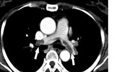
When an Embolus from a deep vein thrombosis reaches the lung, after traversing the right side of the heart, it is trapped in the small arterioles in the lung and is referred to as a Pulmonary Embolus (PE). This is fortuitous, because, although it causes havoc in the lung, it is precluded from reaching the left side of the heart, from where it can be extruded into the general arterial circulation, where it may end up in the brain, resulting in a stroke, or other organ of significance. The lung is a large resilient organ, and can take the embolic abuse better than most other organs.
When an Embolus from a deep vein thrombosis reaches the lung, after traversing the right side of the heart, it is trapped in the small arterioles in the lung and is referred to as a Pulmonary Embolus (PE). This is fortuitous, because, although it causes havoc in the lung, it is precluded from reaching the left side of the heart, from where it can be extruded into the general arterial circulation, where it may end up in the brain, resulting in a stroke, or other organ of significance. The lung is a large resilient organ, and can take the embolic abuse better than most other organs. Even emboli can be extruded from the deep veins of the upper extremities, where thrombi may occur in the Axillary or Subclavian veins, which have been subjected to indwelling catheters.
When the embolus blocks up a small arteriole or even a large pulmonary artery, the oxygen within the air sacs (alveoli), is unable to pass through the lung/blood vessel membrane to reach the blood where it is normally carried to the rest of the body. This results in that area affected by the embolic process in being unable to perform the normal oxygen transfer function and if it’s a large enough area, symptoms of oxygen deprivation will occur.
The individual will become Short of Breath. Often pleurisy chest pain will occur (pain on inspiration). Wheezing, chest wall tenderness without prior trauma, new onset arrhythmia, such as atrial fibrillation, and even unexplained shock can occur. Often the symptoms are vague. A friction rub may be detected on physical examination.Chronic recurrent PE, often associated with vague symptoms or asymptomatic, can result in permanent chronic damage to the heart and lung.
Diagnosis:
The signs and symptoms are as aforementioned. Shortness of Breath and Chest Pain are the most common presenting symptoms. Sometimes extreme anxiety and fear of imminent death is expressed. The patient is often pale. The pulse may be fast in an effort to move oxygen containing blood around faster.
Laboratory studies reveal a fall in the partial pressure of oxygen in the blood (PO2). A routine chest X-ray may show absence of lung marking in a section of the picture, because there is no blood flow through the vessels in that region of the lung. A Ventilation-Perfusion Lung Scan is the non -invasive gold standard test for suggesting the presence of a Pulmonary Embolus.
It’s referred to as a VQ scan and is reported as high probability, intermittent probability, and low probability of a PE. Of course, Pulmonary Arterial Angiography is the definitive diagnostic tool to confirm the presence of a PE. Recently, a high-resolution helical Computed Tomographic Angiography has shown excellent consistency in predicting PE’s presence.
Treatment: As with DVT, , anticoagulants, fibrinolytics, and surgery, are the treatments directed at dissolving the clot, preventing propagation of the clot, preventing new clots from breaking off from the DVT and reaching the lung, and in extremely large Pulmonary Emboli, removing the clot (embolism), which is blocking a major Pulmonary Artery. A Greenfield filter placed in the Inferior Vena Cava is instrumental in preventing further emboli from reaching the lung and can be placed non-invasively.
