Magnetic Resonance Imaging or MRI is an imaging technique that utilizes a magnetic field and pulses of radio wave energy to obtain images. It was invented in 1971 by Paul C. Lauterbur , although it was not used to obtain the first clinically useful images of a patient until 1980. It is not a photograph, but a computerized map or image of radio signals emitted by the human body.
MRI has a wide range of applications in the medical field. It is estimated that as of 2014, there were approximately 25,000 MRI scanners in use world-wide. It is a powerful diagnostic tool as the procedure of choice in the visualization of the soft tissues of the body.
MRI may show problems that may not be seen with other imaging techniques like a plain X-Ray. Also, since it does not use radiation, it is often preferable to a CAT scan.
An MRI may also be taken with the utilization of contrast material (dye) injected into the patient to help demarcate and delineate tissues.
From a safety standpoint, MRI is a safe imaging technique. However, there are certain contraindications to using MRI, including the use in some patients who have implanted metal medical devices like cochlear implants and most permanent pacemakers. Also, since the magnetic field is very strong, there is also a risk of projectile effect where metal objects fly through the air like missiles toward the magnet. Thus, jewelry and other ferromagnetic objects must not be used in the close vicinity of an ongoing MRI scan.
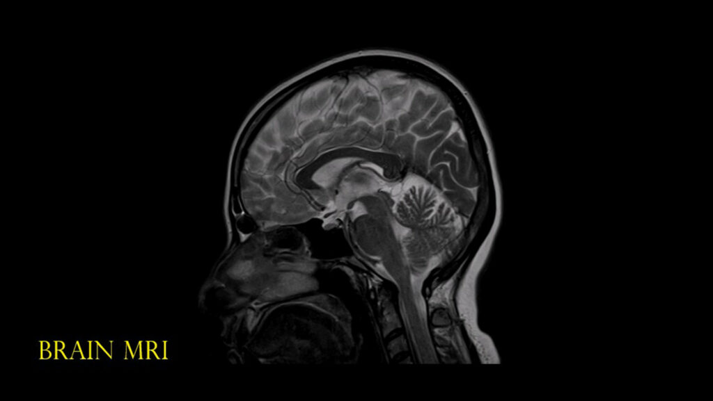
MRI has proven to be the imaging technique of choice in the field of Neurology and Neuroimaging. The contrast between the grey and white matter makes it particularly useful for diseases of the central nervous system. It is also the investigative tool of choice in most neurological cancers.